


Tooth imaging
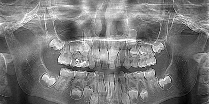
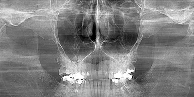
Tooth imaging – the X-ray is a very important auxiliary diagnostic tool that has been used in medicine and dentistry for decades. The modern (digital) approach that meets the needs of contemporary dental diagnostics has become a reality in our country, too.
Advantages of digital radiography:
- Irradiation reduced by 50 – 90%
- Sharper and more precise image
- No chemicals used for imaging
- Reduced development time, therefore, the patient spends less time at the Clinic
- Permanent filing option
- Foil, photo-paper or CD can be the X-ray image medium
- Computer manipulation in the region of interest by the therapist, if the image is stored on a CD
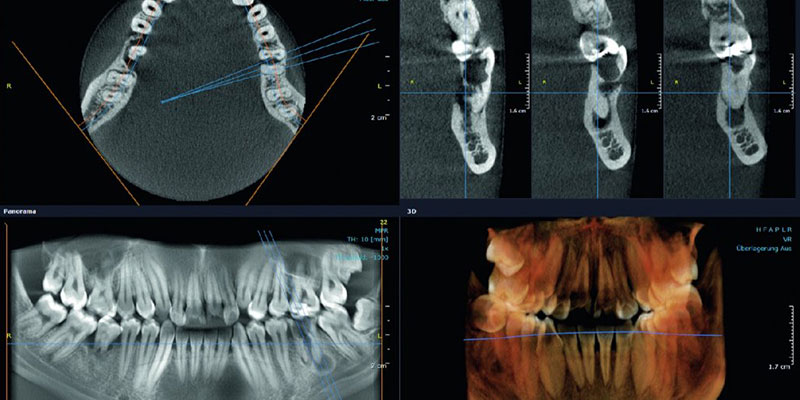
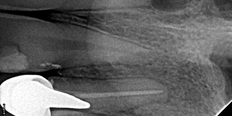
- Measuring different anatomical structures in real proportions: alveolar ridge width, alveolar ridge height, tooth root canal length, cyst or tumour size, etc.
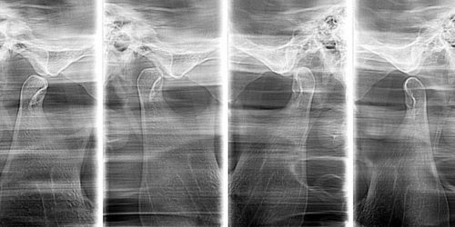
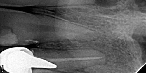
Possible imaging techniques


There are numerous advantages to digital radiology, obviously, but, respecting the preferences and habits of a certain number of dentists, tooth imaging can also be performed using the standard method, on classical X-ray film!
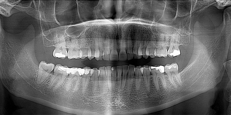
The digital panoramic imaging methods and 3D diagnostics machine is a Kawo OP 3D pro tomograph.
Every patient is protected against X-rays using impregnated clothes that meet the local regulations, as well as by using the appropriate infection control procedures.


